
stories
Feb 28, 2024Support OVC Pet Trust at the 14th Annual OTS Dog Jog
The 2024 OTS Dog Jog will take place on Saturday, March 23, 2024 at the University of Guelph Arboretum. Join us for this family friendly event in support of OVC Pet Trust!

OVC Pet Trust is Canada’s first charitable fund dedicated to improving and advancing companion animal health and well-being. We support innovative discoveries, education and health care that improve the prevention, diagnosis and treatment of diseases of pets.
stories
Feb 28, 2024The 2024 OTS Dog Jog will take place on Saturday, March 23, 2024 at the University of Guelph Arboretum. Join us for this family friendly event in support of OVC Pet Trust!
stories
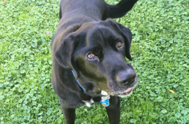
Nov 24, 2023During Pet Cancer Awareness Month, we honour Murphy, a dog whose incredible journey with cancer brought him from Buffalo, New York to the Ontario Veterinary College (OVC) at the University of Guelph.
news
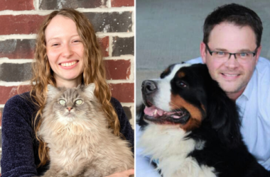
Oct 19, 2023Research shows that a cat or dog that maintains its ideal body weight is less likely to experience chronic ailments such as diabetes, joint or mobility issues and may have a longer lifespan compare